


BRAIN & SPINAL CORD AVM/AVF EMBOLIZATION
Understanding AVM/AVF
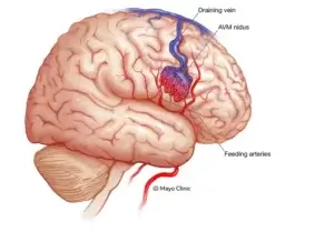
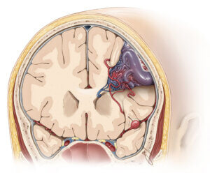
Arteriovenous malformations (AVMs) and arteriovenous fistulas (AVFs) are abnormal connections between arteries and veins in the brain or spinal cord. Normally, blood travels from arteries to capillaries, then to veins. In AVM/AVF, there’s a direct connection, bypassing the capillaries. This disrupts normal blood flow and can lead to serious complications like bleeding (hemorrhage) or stroke.
- Brain AVM/AVF: These are abnormal connections within the brain.
- Spinal Cord AVM/AVF: These occur within or around the spinal cord.
Embolization Procedure
Embolization is a minimally invasive procedure used to treat AVM/AVF and reduce the risk of complications. It’s performed by an interventional radiologist (IR doctor) using X-ray guidance. Here’s a general overview:
- Access: A small incision is made in the groin area.
- Catheter Insertion: A thin, flexible tube (catheter) is inserted into the artery and threaded towards the blood vessels feeding the AVM/AVF.
- Embolic Material Deployment: Once positioned, tiny particles, coils, or a glue-like substance are released through the catheter to block the abnormal connection and reduce blood flow to the AVM/AVF.
- Closure: The catheter is removed, and the incision site is closed.
Benefits of Embolization
Embolization offers several advantages over traditional surgery:
- Minimally invasive
- Less scarring
- Quicker recovery time
- Can be used for AVMs/AVFs in locations difficult to reach surgically
IR Treatment and Embolization
Interventional radiology (IR) plays a crucial role in AVM/AVF embolization. IR doctors are specially trained physicians who use image-guided minimally invasive procedures to diagnose and treat various conditions, including vascular malformations. Their expertise in using X-ray, fluoroscopy, and other imaging techniques allows for precise navigation during embolization.
Here’s how IR is vital in embolization:
- Imaging Guidance: IR doctors utilize real-time imaging to visualize the blood vessels, access the AVM/AVF, and ensure accurate placement of embolic materials.
- Catheter Selection and Navigation: They choose the appropriate catheter size and navigate it through the complex vascular network to reach the target area.
- Embolic Material Selection: IR doctors select the most suitable embolic material (particles, coils, glue) depending on the specific AVM/AVF characteristics.
- Complication Management: They are equipped to handle any potential complications that may arise during the procedure.
Important to Note
- Embolization may not be suitable for all AVM/AVF cases. Sometimes, surgery or a combination of embolization and surgery might be necessary.
- The success rate of embolization depends on various factors, including the size and location of the AVM/AVF.
- Consultation with a qualified neurointerventional radiologist or interventional radiologist is essential to determine the best course of treatment for brain or spinal cord AVM/AVF.
In conclusion, embolization is a valuable tool in the interventional radiologist’s arsenal for treating brain and spinal cord AVM/AVF. It offers a minimally invasive approach to reduce the risk of bleeding and other complications associated with these vascular malformations.